Loculated Pleural Effusion Cxr : Disease Of The Pleura Radiology Key / e intrinsic characteristics of an effusion and its.. Pleural effusions occur as a result of increased fluid formation and/or reduced fluid resorption. Pleural effusions are a common medical problem with more than 50 recognised causes including disease local to the pleura or underlying lung, systemic conditions, organ dysfunction and drugs.1. Treatment depends on the cause. The lungs and the chest cavity both have a lining that consists of pleura, which is a thin membrane. Differentiation of loculated effusions from solid masses.
Differential Diagnosis Of Pleural Effusion from ddxof.com Pleural effusion is a condition in which excess fluid builds around the lung. Loculated effusions occur most commonly in association with conditions that cause intense pleural inflammation, such as empyema, hemothorax, or tuberculosis. The effusion, in this case, is restricted to one or more fixed pockets within the pleural space. Empyema is defined as the presence of pus in the pleural space. no change in position of effusion withchange in position of chest. Pleural effusions are diagnosed in about 1.5 million individuals in the united states annually. Approximately 1 million people develop this abnormality each year in the united states. Meniscus sign is a rim of fluid ascending the lateral chest wall.
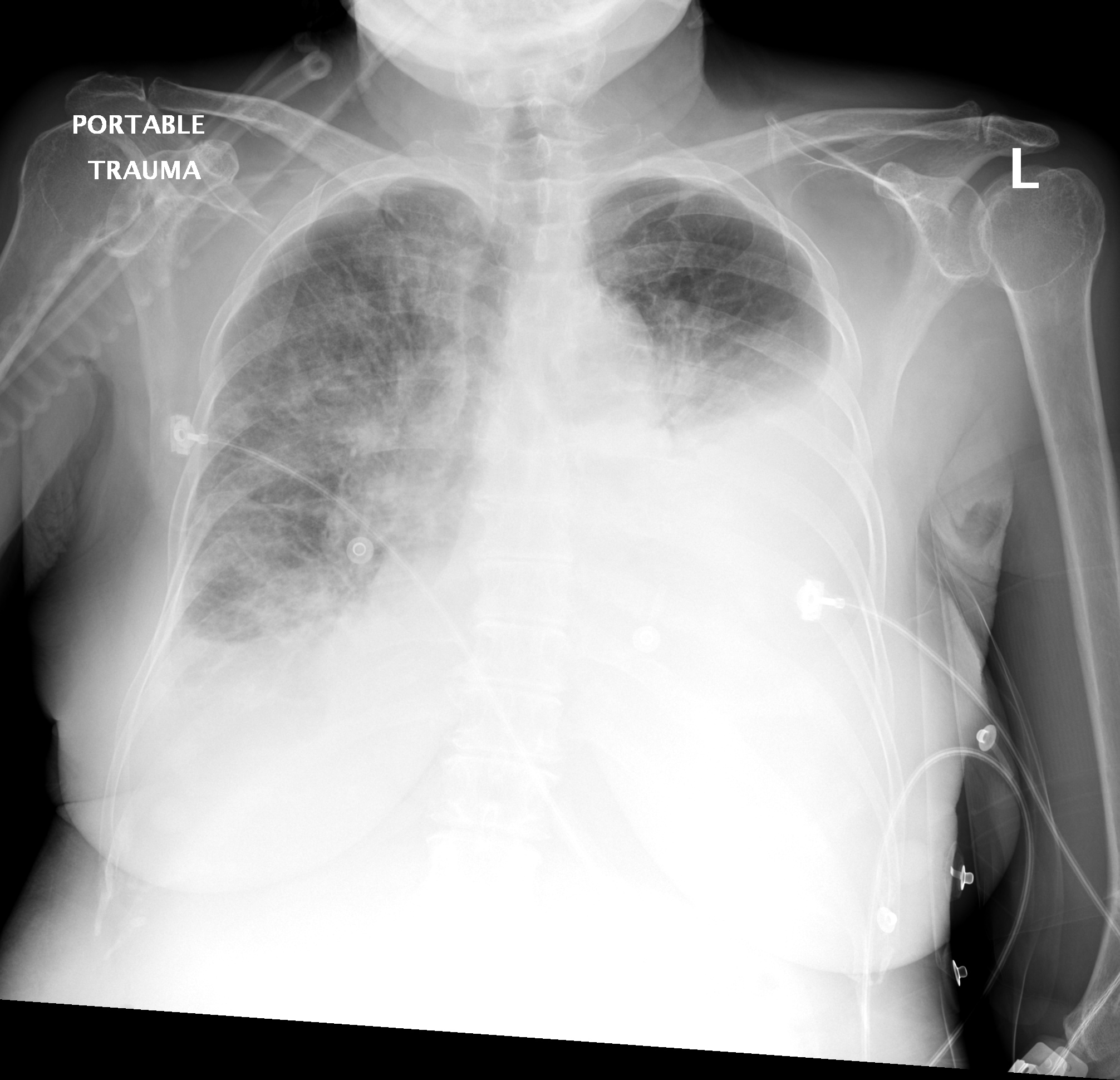
Obliteration of left costophrenic angle with a wide pleural based dome shaped opacity projecting into the lung noted tracking along the cardiophrenic angle and lateral chest wall suggestive of loculated pleural effusion, however the.
Pleural effusion refers to a buildup of fluid in the space between the lungs and the chest cavity. Accompanying adhesions can be identified. What does pleural effusion mean? Empyema is defined as the presence of pus in the pleural space. The underlying lung shrinks and atelectasis develops in a round configuration. Dr bhatia discussing on pleural effusion in #lastminuterevisionpointdiscussionseries. e intrinsic characteristics of an effusion and its. Pleural fluid/serum ldh ratio >0.6. More than one half of these massive pleural effusions are caused by malignancy; produced at parietal and resorbed atvisceral pleura. There is a large left pleural effusion obscuring the lower half of the left hemi thorax. There is always a small amount of fluid around the lung t. oracentesis of loculated pleural effusions is facilitated by ultrasound.
Learn about pleural effusion including causes of pleural effusion. Parapneumonic effusion is a pleural fluid ap/pa cxr: Pleural fluid/serum protein ratio >0.5. What are the pulmonary findings? 9 633 просмотра 9,6 тыс.
Pleural Effusion Work Up Youtube from i.ytimg.com Loculated effusions occur most commonly in association with conditions that cause intense pleural inflammation, such as empyema, hemothorax, or tuberculosis. Empyema is defined as the presence of pus in the pleural space. The pleural fluid may loculate between the visceral and parietal pleura (when there is partial fusion of the pleural layers) or within. Accompanying adhesions can be identified. Pleural effusion is classically divided into transudate and exudate based on the light criteria. no change in position of effusion withchange in position of chest. Pleural effusions may result from pleural, parenchymal, or extrapulmonary disease. Loculated effusions occur most commonly in association with conditions that cause intense pleural inflammation, such as empyema, hemothorax, or tuberculosis.
Determine if it can be tapped.
Blunting of costophrenic angle initially. Other causes are complicated parapneumonic effusion. Parapneumonic effusion is a pleural fluid ap/pa cxr: A pleural effusion is accumulation of excessive fluid in the pleural space, the potential space that surrounds each lung. Bhatia medical coaching institute, dbmci. Approximately 1 million people develop this abnormality each year in the united states. Differentiation of loculated effusions from solid masses. Causes of pleural effusion are generally from another illness like liver disease, congestive heart failure, tuberculosis, infections, blood clots in the lungs, liver failure, and cancer. The underlying lung shrinks and atelectasis develops in a round configuration. The cardiac silhouette is also obscured. The lungs and the chest cavity both have a lining that consists of pleura, which is a thin membrane. My pleural effusion healed without treatment. Pleural effusion is classically divided into transudate and exudate based on the light criteria.
9 633 просмотра 9,6 тыс. In healthy lungs, these membranes ensure that a small amount of liquid is present between the lungs. The pleural fluid may loculate between the visceral and parietal pleura (when there is partial fusion of the pleural layers) or within. The underlying lung shrinks and atelectasis develops in a round configuration. My pleural effusion healed without treatment.
Pleural Effusion Dr Mahesh from cdn.slidesharecdn.com Pleural effusions may result from pleural, parenchymal, or extrapulmonary disease. Differentiation of loculated effusions from solid masses. Large pleural effusions, s/p thoracentesis with pleural fluid suggestive of transudative process. It is commonly known as water on the lungs. Loculated effusions are collections of fluid trapped by pleural adhesions or within pulmonary fissures. There is always a small amount of fluid around the lung t. Occasionally, a focal intrafissural fluid collection may look like a lung mass. 9 633 просмотра 9,6 тыс.
Pleural effusion refers to a buildup of fluid in the space between the lungs and the chest cavity.
Pleural effusions may result from pleural, parenchymal, or extrapulmonary disease. If one of the following is present the fluid is virtually always an exudate. Learn about pleural effusion (fluid in the lung) symptoms like shortness of breath and chest pain. Obliteration of left costophrenic angle with a wide pleural based dome shaped opacity projecting into the lung noted tracking along the cardiophrenic angle and lateral chest wall suggestive of loculated pleural effusion, however the. Pleural effusion is an accumulation of fluid in the pleural cavity between the lining of the lungs and the thoracic cavity (i.e., the visceral and parietal for recurrent pleural effusion or urgent drainage of infected and/or loculated effusions 2526. Loculated pleural effusion on cxr. 9 633 просмотра 9,6 тыс. Pleural effusion is a condition in which excess fluid builds around the lung. This situation most commonly is seen in patients with heart failure. The underlying lung shrinks and atelectasis develops in a round configuration. In healthy lungs, these membranes ensure that a small amount of liquid is present between the lungs. Transudates or exudates as defined by lights criteria. Differentiation of loculated effusions from solid masses.


0 Komentar